
Induction of mesenchymal stem cell chondrogenesis by polyacrylate substrates*
Cell Specific – MSCs
(In association with University of Liverpool)
Background
Many studies have explored the potential of different stem cell types, such as MSCs to generate chondrocytes in vitro, the long-term aim being to determine if stem cell-derived chondrocytes have the potential to repair osteoarthritic lesions. In order to develop culture conditions to direct stem cells to differentiate to chondrocytes in vitro, it is important to understand the mechanisms that regulate the differentiation of these cells in vivo. Using this understanding will lead to the possibility of designing substrates that promote differentiation down this route.
Challenge
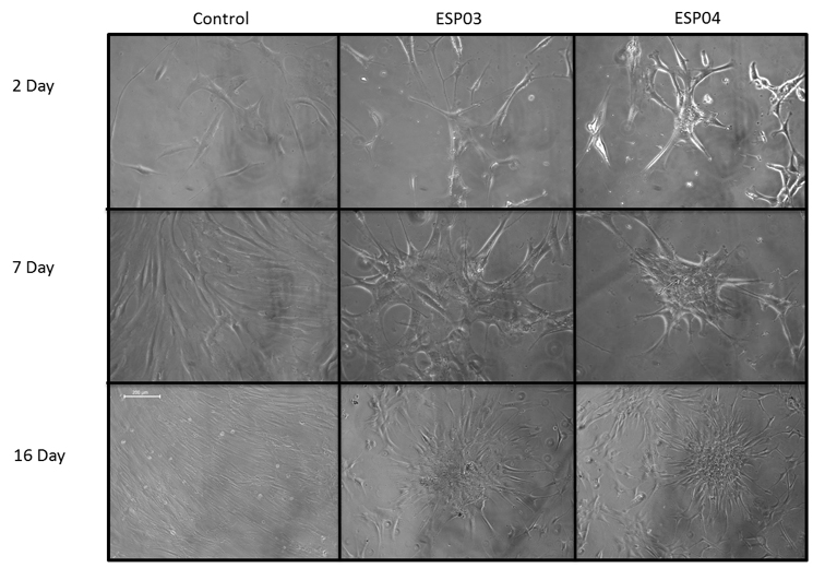
Mesenchymal stem cells (MSCs) can generate chondrocytes in vitro, but typically need to be cultured as aggregates in the presence of transforming growth factor beta (TGF-b), which makes scale-up difficult.
Here we investigated if polyacrylate substrates modelled on the functional group composition and distribution of the Arg-Gly-Asp (RGD) integrin-binding site could induce MSCs to undergo chondrogenesis in the absence of exogenous TGF-b.
Study
The candidate substrate designs were based upon complex multi-monomeric, acrylic based polymers, each monomeric unit containing specific functional groups, the composition and distribution of which can be discretely modified in terms of the starting monomer ratio in order to tailor the surface properties. The polymers are synthesised by free radical polymerisation using proprietary controlled process techniques to ensure that the functional group chemistries of the constituent monomers are evenly distributed throughout the polymeric backbone. The composition and distribution of the functional group chemistries along the polymeric backbone influence the charge, charge density, hydrophobic/hydrophilic balance and surface stereochemistry of the resultant coating substrate.
R1 and R2 denote interchangeable side groups.

Chemical structure of acrylates materials. Acrylate monomer and poly-acrylate formulae are shown. R1 and R2 denote potential side groups.
Results
Within a few days of culture on the biomimetic polyacrylates, both mouse and human MSCs, and a mesenchymal-like mouse-kidney-derived stem cell line, began to form multi-layered aggregates and started to express the chondrocyte-specific markers, Sox9, collagen II and aggrecan. Moreover, collagen II tended to be expressed in the centre of the aggregates, similarly to developing limb buds in vivo.
Surface analysis of the substrates indicated that those with the highest surface amine content were most effective at promoting MSC chondrogenesis.
These results highlight the importance of surface group functionality and the distribution of those groups in the design of substrates to induce MSC chondrogenesis.
*Acta biomaterialia 9(4) · December 2012
![]()
